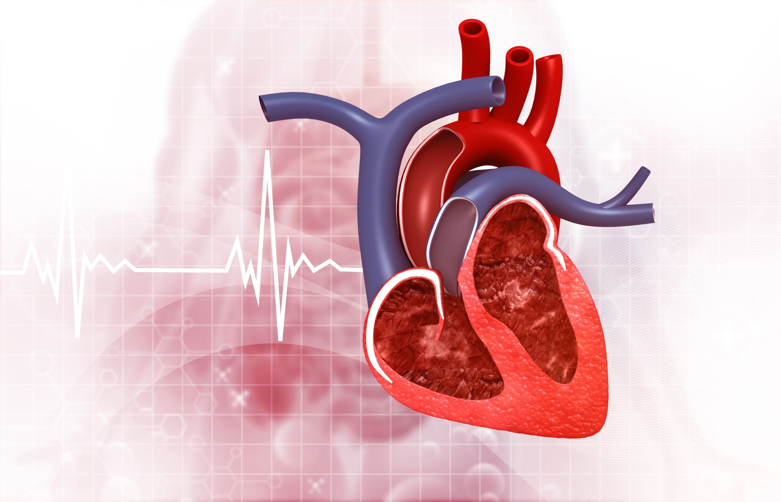
Cardiac Sciences Centre
The Kislay Iconic & Batra Hospital Heart Centre represents a groundbreaking collaboration, combining Kislay Iconic's innovative healthcare solutions with Batra Hospital's renowned clinical expertise. Located in the heart of Delhi, this state-of-the-art facility is dedicated to providing comprehensive and compassionate
Non-invasive Diagnostics
Interventional Cardiology
Interventional Cardiology

Advanced echocardiography, stress testing, and Holter monitoring.
Interventional Cardiology
Interventional Cardiology
Interventional Cardiology

Coronary angioplasty, stenting, and pacemaker implantation.
Cardiothoracic Surgery
Cardiothoracic Surgery
Cardiothoracic Surgery

Bypass surgery, valve repair/replacement, and a range of other surgical procedures.
Cardiac Rehabilitation
Cardiothoracic Surgery
Cardiothoracic Surgery

A dedicated program to help patients recover and maintain heart health after a cardiac event.
Cardiac Sciences Centre
Coronary Artery Diseases
Coronary Artery Diseases
Coronary Artery Diseases

The coronary arteries, which carry blood to your heart, are impacted by coronary artery disease (CAD). One or more of your coronary arteries get blocked or progressively narrow with CAD. The most frequent symptom is angina, which causes chest pain. A heart attack or other consequences like arrhythmia or heart failure can result from CAD.
Heart Attack
Coronary Artery Diseases
Coronary Artery Diseases

A myocardial infarction, often known as a heart attack, is a very hazardous ailment that develops when the blood supply to your heart muscle is cut off. Although numerous potential causes exist, a blockage in one or more of your heart's arteries is the most common. The injured cardiac muscle will start to deteriorate without blood flow. A heart attack might result in lasting cardiac damage and perhaps death if blood flow isn't rapidly restored.
Heart Failure
Coronary Artery Diseases
Heart Valve Disease

Heart failure is a chronic disorder in which your heart cannot continuously pump blood efficiently enough to meet your body's demands. Exercise, medication, and maybe surgery are all part of the course of treatment for heart failure. One aspect that affects your outlook is how well you take care of yourself.
Heart Valve Disease
Congenital Heart Disease/Pediatric Cardiology
Heart Valve Disease

Any of a number of disorders that impair the proper function of one or more of your heart's valves is referred to as heart valve disease. Heart valve problems can make your heart work harder if it is not treated. Your quality of life may be negatively impacted, and it can even endanger your life. Your healthcare practitioner may be able to replace or repair your heart valves by surgery or minimally invasive treatment in many circumstances, restoring normal function and enabling you to resume your normal activities.
Congenital Heart Disease/Pediatric Cardiology
Congenital Heart Disease/Pediatric Cardiology
Congenital Heart Disease/Pediatric Cardiology

Congenital heart disease (CHD) is a structural flaw or issue with the heart that exists from birth. Examples include:
a crack in the heart's lining.
complications with the blood vessels (too many or too few, blood flowing too slowly, to the wrong place or in the wrong direction).
the cardiac valves are malfunctioning.
While some CHD cases are minor and may not manifest any symptoms, others can be fatal and necessitate medical attention.
Early heart problem detection is possible (before a baby is born or shortly afterwards). However, in other cases, it takes until childhood, adolescence, or adulthood to diagnose CHD.
Aortic Aneurysm
Congenital Heart Disease/Pediatric Cardiology
Congenital Heart Disease/Pediatric Cardiology

The aorta is the body's biggest artery. It transfers oxygen-rich blood from your heart to the rest of your body. It has a curled candy cane form. Your heart's aorta ascends from there. Your abdomen is the last destination of the descending aorta (belly).
Any artery can become a target for an aneurysm. When the wall of your aorta weakens, an aortic aneurysm can form. The weak spot of your aorta develops a balloon-like protrusion due to the pressure of blood pumping through the artery. An aortic aneurysm is the name given to this protrusion.
Transient Ischemic Attack
Transient Ischemic Attack
Transient Ischemic Attack

A transient ischemic attack (TIA) is a temporary period of symptoms similar to those of a stroke. A TIA usually lasts only a few minutes and doesn't cause permanent damage. Often called a ministroke, a TIA may be a warning. About 1 in 3 people who have a TIA will eventually have a stroke, with about half occurring within a year after the TIA. A TIA can serve as both a warning of a future stroke and an opportunity to prevent it.
Long QT Syndrome (LQTS)
Transient Ischemic Attack
Transient Ischemic Attack

An electrical condition of the heart is known as long Q-T syndrome. The movement of ions (electrically charged particles of sodium, calcium, potassium, and chloride) into and out of the cardiac cells causes the heart's electrical activity. This flow is regulated by tiny ion channels. The ion channel deficiency that results in LQTS prolongs the time it takes for the electrical system to recharge after each heartbeat. The risk of torsade de pointes, a fatal variation of ventricular tachycardia, increases when the Q-T interval is longer than usual.
Heart Block
Transient Ischemic Attack
Heart Block

When the electrical signal that regulates your heartbeat is partially or totally blocked, it is known as a heart block or an AV block. As a result, your heart beats more slowly or skips beats, making it unable to pump blood efficiently. Dizziness, fainting, exhaustion, and shortness of breath are some of the symptoms. Implanting a pacemaker is a common treatment.
HOW IT WORKS
VIDEO CONSULTATION WITH A DOCTOR
MEDICAL VISA FOR TREATMENT IN INDIA
MEDICAL VISA FOR TREATMENT IN INDIA

MEDICAL VISA FOR TREATMENT IN INDIA
MEDICAL VISA FOR TREATMENT IN INDIA
MEDICAL VISA FOR TREATMENT IN INDIA

COMPLETE TREATMENT & SURVIVAL
MEDICAL VISA FOR TREATMENT IN INDIA
COMPLETE TREATMENT & SURVIVAL

MEET OUR DOCTORS

Prof. (Dr.) Upendra Kaul
Prof. (Dr.) Upendra Kaul
Prof. (Dr.) Upendra Kaul
Qualification: MD, DM, FCSI, FICC, FACC, FAPSIC, FSCAI, FAMS
Designation: Chairman, Batra Heart Centre

Dr. R.D. Yadave
Prof. (Dr.) Upendra Kaul
Prof. (Dr.) Upendra Kaul
Qualification: MD, DM (Cardiology)
Designation: Senior Consultant, Interventional Cardiology and Head of the Cath Lab

Dr. Sanjiv Sharma
Prof. (Dr.) Upendra Kaul
Dr. Sanjiv Sharma
Qualification: MD (Medicine), DM (Cardiology)
Designation: HOD, Interventional Cardiology

Dr. Varsha Koul
Dr. Sanjay Kumar Pandey
Dr. Sanjiv Sharma
Qualification: MBBS, MD (Internal Medicine), DNB (Cardiology), MNAMS
Designation: Senior Consultant & Coordinator, Dept of Cardiology

Dr. Sanjay Kumar Pandey
Dr. Sanjay Kumar Pandey
Dr. Sanjay Kumar Pandey
Qualification: MS, MCh (CTVS)
Designation: Chief Cardiac Surgeon & HOD CTVS

Dr Pawan Zutshi
Dr. Sanjay Kumar Pandey
Dr. Sanjay Kumar Pandey
Qualification: MBBS, MD (Internal Medicine), DNB (Cardiology), FSCAI
Designation: Senior Consultant, Cardiology
TREATMENTS
Intra-arterial Vasodilation
Intra-arterial Vasodilation
Intra-arterial Vasodilation
One of the suggested therapies to lessen the effects of cerebral vasospasm brought on by aneurysmal subarachnoid haemorrhage and resistance to normal management is intra-arterial (IA) vasodilator therapy. Its usefulness and effectiveness, however, are not well established.
External Counter Pulsation
Intra-arterial Vasodilation
Intra-arterial Vasodilation
Chest pain treatment involves enhanced external counterpulsation (EECP) therapy. It treats chronic stable angina, long-term chest pressure or pain that doesn't respond to conventional treatments. Some patients who require a method to restore blood flow to their heart but are ineligible for surgery may also be advised to undergo EECP therapy. The EECP therapy increases your blood flow by applying pressure. It is an outpatient, non-invasive procedure. The treatment usually lasts for seven weeks.
Electrocardiogram(EKG)
Intra-arterial Vasodilation
Electrocardiogram(EKG)
Temporary electrodes are placed on your chest and limbs during an electrocardiogram (EKG/ECG), which records, tracks, and records the electrical activity of your heart (which regulates your heartbeats) for diagnostic purposes. A computer converts the data into a wave pattern your healthcare physician can understand. This is a short, painless test that is not intrusive. As part of a stress test, an EKG can be obtained while resting or working out.
Echocardiogram(ECHO)
Coronary Computed Tomography Angiogram(CTA)
Electrocardiogram(EKG)
An echocardiogram (echo) is a visual representation of the beats of your heart. Your doctor will perform an echo test on you to take images of your heart's chambers and valves using ultrasonography (high-frequency sound waves) from a hand-held wand inserted into your chest. This aids the medical professional in assessing how well your heart is pumping. Medical professionals frequently mix echo with Doppler ultrasound and colour Doppler procedures to assess blood flow across your heart's valves. No radiation is used in echocardiography. This distinguishes an echo from other examinations, such as X-rays and CT scans, which employ negligible radiation.
Coronary Computed Tomography Angiogram(CTA)
Coronary Computed Tomography Angiogram(CTA)
Coronary Computed Tomography Angiogram(CTA)
A CT angiography is a test that allows doctors to see your blood arteries and tissues. Your doctor injects a dye that highlights your blood vessels and tissues during the test. The highlighted areas are then examined using a CT scan, a type of specialist X-ray.
Cardiac Magnetic Resonance Imaging (MRI)
Coronary Computed Tomography Angiogram(CTA)
Coronary Computed Tomography Angiogram(CTA)
Heart MRIs use radio waves and magnets to make images of your heart, with nothing you can see or feel entering your body. The components of your heart, including its chambers, valves, and muscles, as well as how effectively they are functioning and how your blood is moving, can be seen on a cardiac MRI. Your healthcare professional can diagnose the issue and determine what's wrong thanks to these finely detailed, high-quality photos in two or three dimensions.
Positron Emission Tomography Scan(PET)
Positron Emission Tomography Scan(PET)
Positron Emission Tomography Scan(PET)
An imaging test called a positron emission tomography (PET) scan creates images of your organs and tissues. The test uses a PET scanner and a radioactive substance that is safe to inject called a radiotracer. When sick cells absorb a lot of the radiotracer, the scanner picks this up as a possible health issue.
Cardiac Catheterization
Positron Emission Tomography Scan(PET)
Positron Emission Tomography Scan(PET)
Cardiac catheterization, often known as cardiac cath or coronary angiography, is an invasive imaging technique that lets your doctor assess how well your heart is working. To access your coronary arteries, your healthcare professional inserts a catheter (a thin tube) into a blood artery in your arm or groyne. Heart catheterizations are occasionally used in conjunction with other procedures to diagnose and treat issues. For instance, a doctor could replace a damaged heart valve or correct a birth defect in your heart without performing traditional surgery or making a significant incision.
Coronary Artery Bypass Surgery
Positron Emission Tomography Scan(PET)
Coronary Artery Bypass Surgery
Coronary artery bypass grafting is a surgery that restores blood flow to areas of your heart that aren’t getting enough blood. This surgery can improve your heart function and how you feel, especially when you’ve just had a heart attack or there’s an increased risk of having one shortly.
Copyright © 2024 KISLAY Iconic HealthCare Pvt Ltd - All Rights Reserved.
Powered by Kislay Group
